|
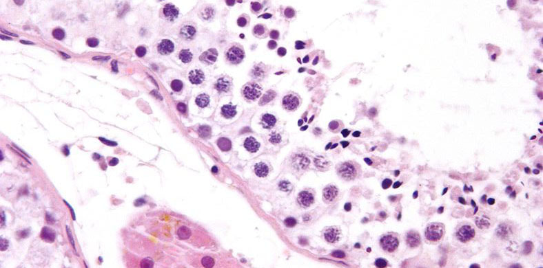
(Image courtesy of Nephron, CC BY-SA 3.0) Seminiferous Tubules are located in the testicles, the two oval-shaped organs on either side of a male’s penis. There are around 800 seminiferous tubules in each testicle, and this is where meiosis and subsequent development of spermatozoa occurs. In a mature adult male, each of these tubules creates thousands of sperm every second. The tubules are tightly looped throughout the testes, and are incredibly long: perhaps as much as a mile between the two testes! Their tissue is lined with Sertoli Cells, a specialized cell type that is responsible for creating new sperm. They do so by secreting a protein that increases the concentration of testosterone inside the seminiferous tubules. There are two distinct types of seminiferous tubules:
It is important to note that sperm are not entirely complete in their maturation upon leaving the seminiferous tubules, and will undergo further maturation in the epididymis, such as developing the tails that give them their motility. Nuts & Bolts: What are Seminiferous Tubules? To learn more about, please visit our series of posts about male reproduction and contraception: Key terms: Meiosis - a type of cell division that results in four daughter cells each with half the number of chromosomes of the parent cell, as in the production of gametes and plant spores. Penis - the male genital organ of higher vertebrates, carrying the duct for the transfer of sperm during copulation. In humans and most other mammals, it consists largely of erectile tissue and serves also for the elimination of urine. Protein - essential nutrients for the human body that are one of the building blocks of body tissue and can also serve as a fuel source. Testicle - an organ which produces spermatozoa (male reproductive cells). Testosterone - the primary sex hormone and anabolic steroid in males, responsible for the development of male reproductive tissues and secondary sexual characteristics. For additional terminology related to male contraception and the male reproductive system, please visit our glossary: Sources/References: Mahfouz R, Sharma R, Thiyagarajan A, Kale V, Gupta S, Sabanegh E, Agarwal A (2010). "Semen characteristics and sperm DNA fragmentation in infertile men with low and high levels of seminal reactive oxygen species". Fertil. Steril. 94 (6): 2141–6. doi:10.1016/j.fertnstert.2009.12.030 Gunes S, Al-Sadaan M, Agarwal A (2015). "Spermatogenesis, DNA damage and DNA repair mechanisms in male infertility". Reprod. Biomed. Online. 31 (3): 309–19. doi:10.1016/j.rbmo.2015.06.010 Nakata H. Morphology of mouse seminiferous tubules. Anat Sci Int. 2019 Jan;94(1):1-10. doi: 10.1007/s12565-018-0455-9. Epub 2018 Aug 20. Mendez JA, Emery JL. The seminiferous tubules of the testis before, around and after birth. J Anat. 1979 May; 128(Pt 3):601-7. Wing TY, Christensen AK. Morphometric studies on rat seminiferous tubules. Am J Anat. 1982 Sep;165(1):13-25. doi: 10.1002/aja.1001650103. Yoshida S. From cyst to tubule: innovations in vertebrate spermatogenesis. Wiley Interdiscip Rev Dev Biol. 2016 Jan-Feb;5(1):119-31. doi: 10.1002/wdev.204. Epub 2015 Aug 25. For additional publications related to male contraception and the male reproductive system, please visit our publications page:
Comments are closed.
|
Categories
All
Archives
June 2024
|
|
|
Donate to Male Contraceptive InitiativeYour generous donation makes a difference!
|
© Male Contraceptive Initiative. All rights reserved.


 RSS Feed
